|
|||||||||||||
 |
|||||||||||||
X-Ray Inspection
|
|||||||||||||
X-ray inspection capability is a vital tool not just in medicine but in industry. During my tenure with a major cellphone power amplifier company as an RF Applications Engineer, I relied heavily on x-ray imaging for many of the teardown reports that I wrote on competitors' products. It included everything from determining metal layers in integrated circuits to deducing multi-layer ceramic PCB routing lines and distributed components to just getting a quick look inside a shielded enclosure without needing to disassemble it. A skilled x-ray technician can make a huge difference in the quality of information that can be gleaned from the images since angle, power level, and focusing takes a deft touch. My first experience with using x-rays for inspection was while doing automated test system design for production base station equipment companies. Very stringent PIM (passive intermodulation) specifications were required for high power filters in order to minimize 3rd and higher level intermods. The N-type connectors ended up being the Achilles' heel of the switching filter matrix, and we had ordered a large quantity from one highly qualified manufacturer. Quite a few needed to be returned to the factory for rework because of failed intermod tests. The relationship got a bit contentious after a while. One problem ended up being that the connectors were supposed to have staking pins to guarantee that the connector body could not turn during torqueing. The vendor swore to me that their inspection process was so perfect that there was no way any could have been missed. Fortunately for me, the company I worked for also happened to manufacture x-ray tubes and had an x-ray machine for testing the tubes. I had the production test technician slide one of my suspect filters into the machine and sure enough, the filters that had the stainless steel pins installed showed in high contrast to the aluminum filter cases. I then had a way to verify every filter, which was not possible with only a visual inspection because an epoxy backfill material was placed into the staking pin hole. X-Ray Inspection
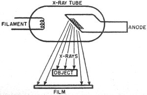
Penetrating powers of x-rays help industry probe secrets of materials and new products. X-rays are similar to radio waves in that they are electronically produced, are invisible, and travel at the speed of light. X-rays, however, are in the extremely high frequency range and have very short wave-lengths. Even the shortest of radio waves, the so-called "microwaves" - are gigantic by comparison. A typical wavelength of x-ray radiation is 0.0000000001 meter (one ten-billionth of a meter). These extremely short wavelengths have great penetrating power, and can pass through substances which light cannot penetrate. Besides their well-known use in dental and medical examinations, x-rays have many industrial applications. For example, x-ray apparatus is used to detect internal flaws in metal castings, check packaged foods for presence of foreign particles, inspect welds, check the alignment of elements in electron tubes, check the centering of the wire in insulated cables, etc. Anode of this gigantic x-ray unit gets one million volts. G-E built it for Sutter Hospital, Sacramento, California. As shown in the diagram, the x-ray tube is basically a diode. Electrons emitted from the filament are attracted down the length of the tube to the copper anode. The anode contains a tungsten insert which acts as a target for the electrons. Traveling at a very high speed, the electrons strike the target, producing the x-rays. To give the electron stream its high velocity, a large amount of voltage must be applied to the tube. Plate voltages as high as two million volts have been used in commercial x-ray units.
Electrons from filament hit anode at high speed; resultant radiation creates x-rays. The object to be x-rayed is placed between the x-ray tube and a sheet of photosensitive film. The x-rays penetrate the object and strike the film. This action produces a shadow image of the internal structure of the object. For example, if the object being examined is a pulley belt with internal reinforcement wires, the x-rays will pass easily through the rubber portion of the belt, but will be obstructed by the wires. The developed film will therefore show an image of the wires (see photograph above). In some applications, speed of inspection is an important feature and the time required to develop the film introduces an objectionable delay. In these cases, a fluorescent screen is used instead of the film. Such screens glow where they are exposed to x-rays, and thus produce an immediate image. An installation of this type is known as a fluoroscope.
Posted September 21, 2022 |
|||||||||||||
 |
|||||||||||||
|
|||||||||||||


|
||||||||||||||||||||||||||||||||||||